
15.5: Рибосоми та синтез білка
- Page ID
- 1903

Навички для розвитку
- Опишіть різні етапи синтезу білка
- Обговорити роль рибосом в синтезі білка
Синтез білків споживає більше енергії клітини, ніж будь-який інший метаболічний процес. У свою чергу, білки становлять більшу масу, ніж будь-який інший компонент живих організмів (за винятком води), а білки виконують практично кожну функцію клітини. Процес трансляції, або синтезу білка, передбачає розшифровку повідомлення мРНК в поліпептидний продукт. Амінокислоти ковалентно нанизані разом шляхом переплетення пептидних зв'язків довжиною від приблизно 50 залишків амінокислот до більш ніж 1,000. Кожна окрема амінокислота має аміногрупу (NH 2) і карбоксильну (СООН) групу. Поліпептиди утворюються тоді, коли аміногрупа однієї амінокислоти утворює амідну (тобто пептидну) зв'язок з карбоксильною групою іншої амінокислоти (рис.\(\PageIndex{1}\)). Ця реакція каталізується рибосомами і генерує одну молекулу води.
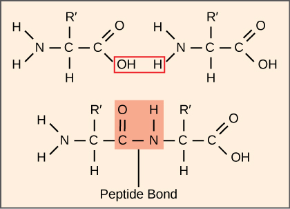
Машини синтезу білка
Крім шаблону мРНК, багато молекули і макромолекули сприяють процесу трансляції. Склад кожного компонента може відрізнятися в різних видах; наприклад, рибосоми можуть складатися з різної кількості РРНК і поліпептидів залежно від організму. Однак загальні структури та функції механізмів синтезу білка можна порівняти від бактерій до клітин людини. Переклад вимагає введення шаблону мРНК, рибосом, тРНК та різних ферментативних факторів.

Рибосоми
Ще до перекладу мРНК клітина повинна інвестувати енергію для побудови кожної своєї рибосом. У кишковій паличці існує від 10 000 до 70 000 рибосом, присутніх у кожній клітині в будь-який момент часу. Рибосома - це складна макромолекула, що складається з структурних та каталітичних РРНК та багатьох різних поліпептидів. У еукаріотів ядерце повністю спеціалізується для синтезу та складання РРНК.
Рибосоми існують в цитоплазмі у прокаріотів і в цитоплазмі і шорсткому ендоплазматичному ретикулумі у еукаріотів. Мітохондрії та хлоропласти також мають власні рибосоми в матриксі та стромі, які виглядають більш схожими на прокаріотичні рибосоми (і мають аналогічну чутливість до ліків), ніж рибосоми безпосередньо поза їх зовнішніми оболонками в цитоплазмі. Рибосоми дисоціюються на великі і малі субодиниці, коли вони не синтезують білки і реассоціюються під час ініціації трансляції. У кишкової палички мала субодиниця описується як 30S, а велика субодиниця - 50S, в цілому 70-ті (нагадаємо, що одиниці Сведберга не є адитивними). Рибосоми ссавців мають невелику субодиницю 40S і велику субодиницю 60S, загалом 80S. Мала субодиниця відповідає за зв'язування шаблону мРНК, тоді як велика субодиниця послідовно зв'язує тРНК. Кожна молекула мРНК одночасно перекладається багатьма рибосомами, всі синтезують білок в одному напрямку: зчитуючи мРНК від 5' до 3' і синтезуючи поліпептид від N кінцевої до закінчення С. Повна структура МРНК/полі-рибосоми називається полісомою.
ТРНК
ТРНК - це структурні молекули РНК, які транскрибували з генів РНК-полімеразою III. Залежно від виду, в цитоплазмі існує від 40 до 60 типів ТРНК. Виконуючи роль адаптерів, специфічні тРНК зв'язуються з послідовностями на шаблоні мРНК і додають відповідну амінокислоту в поліпептидний ланцюг. Тому тРНК - це молекули, які насправді «переводять» мову РНК на мову білків.
З 64 можливих кодонів мРНК - або триплетних комбінацій A, U, G і C - три визначають припинення синтезу білка, а 61 вказують додавання амінокислот до поліпептидного ланцюга. З них 61 один кодон (AUG) також кодує ініціювання перекладу. Кожен тРНК антикодон може базувати пару з одним з кодонів мРНК і додати амінокислоту або припинити трансляцію, відповідно до генетичного коду. Наприклад, якщо послідовність CUA відбулася на шаблоні мРНК у правильному кадрі читання, це зв'яже тРНК, що виражає комплементарну послідовність, GAU, яка була б пов'язана з амінокислотним лейцином.
Як адаптерні молекули перекладу, дивно, що тРНК можуть вмістити стільки специфічності в такий невеликий пакет. Врахуйте, що тРНК повинні взаємодіяти з трьома факторами: 1) вони повинні бути розпізнані правильною аміноацилсинтетазою (див. Нижче); 2) вони повинні бути розпізнані рибосомами; і 3) вони повинні зв'язуватися з правильною послідовністю в мРНК.
Аміноацил-тРНК-синтетази
Процес синтезу пре-тРНК РНК-полімеразою III створює лише частину РНК молекули адаптера. Відповідну амінокислоту необхідно додати пізніше, як тільки тРНК буде оброблена і експортована в цитоплазму. Через процес «зарядки» тРНК кожна молекула тРНК пов'язана зі своєю правильною амінокислотою групою ферментів, званих аміноацильними тРНК синтетазами. Принаймні один тип аміноацильної тРНК синтетази існує для кожної з 20 амінокислот; точна кількість аміноацильних тРНК синтетаз залежить від видів. Ці ферменти спочатку зв'язують і гідролізують АТФ, щоб каталізувати високоенергетичний зв'язок між амінокислотою та аденозинмонофосфатом (AMP); молекула пірофосфату виганяється в цій реакції. Активована амінокислота потім переноситься в тРНК, і AMP вивільняється.
Механізм синтезу білка
Як і при синтезі мРНК, синтез білка можна розділити на три фази: ініціювання, подовження та припинення. Процес перекладу схожий у прокаріотів і еукаріот. Тут ми розглянемо, як відбувається переклад у E. coli, представницького прокаріота, і уточнимо будь-які відмінності між прокаріотичним та еукаріотичним перекладом.
Ініціація перекладу
Синтез білка починається з утворення комплексу ініціації. У кишковій паличці цей комплекс включає невелику рибосому 30S, шаблон мРНК, три фактори ініціації (IF-1, IF-2 та IF-3) та спеціальний ініціатор тРНК, який називається\(\text{tRNA}_\text{f}^\text{Met}\). The initiator tRNA interacts with the start codon AUG (or rarely, GUG), links to a formylated methionine called fMet, and can also bind IF-2. Formylated methionine is inserted by \(\text{fMet} - \text{tRNA}_\text{f}^\text{Met}\) at the beginning of every polypeptide chain synthesized by E. coli, but it is usually clipped off after translation is complete. When an in-frame AUG is encountered during translation elongation, a non-formylated methionine is inserted by a regular Met-tRNAMet.
In E. coli mRNA, a sequence upstream of the first AUG codon, called the Shine-Dalgarno sequence (AGGAGG), interacts with the rRNA molecules that compose the ribosome. This interaction anchors the 30S ribosomal subunit at the correct location on the mRNA template. Guanosine triphosphate (GTP), which is a purine nucleotide triphosphate, acts as an energy source during translation—both at the start of elongation and during the ribosome’s translocation.
In eukaryotes, a similar initiation complex forms, comprising mRNA, the 40S small ribosomal subunit, IFs, and nucleoside triphosphates (GTP and ATP). The charged initiator tRNA, called Met-tRNAi, does not bind fMet in eukaryotes, but is distinct from other Met-tRNAs in that it can bind IFs.
Instead of depositing at the Shine-Dalgarno sequence, the eukaryotic initiation complex recognizes the 7-methylguanosine cap at the 5' end of the mRNA. A cap-binding protein (CBP) and several other IFs assist the movement of the ribosome to the 5' cap. Once at the cap, the initiation complex tracks along the mRNA in the 5' to 3' direction, searching for the AUG start codon. Many eukaryotic mRNAs are translated from the first AUG, but this is not always the case. According to Kozak’s rules, the nucleotides around the AUG indicate whether it is the correct start codon. Kozak’s rules state that the following consensus sequence must appear around the AUG of vertebrate genes: 5'-gccRccAUGG-3'. The R (for purine) indicates a site that can be either A or G, but cannot be C or U. Essentially, the closer the sequence is to this consensus, the higher the efficiency of translation.
Once the appropriate AUG is identified, the other proteins and CBP dissociate, and the 60S subunit binds to the complex of Met-tRNAi, mRNA, and the 40S subunit. This step completes the initiation of translation in eukaryotes.
Translation, Elongation, and Termination
In prokaryotes and eukaryotes, the basics of elongation are the same, so we will review elongation from the perspective of E. coli. The 50S ribosomal subunit of E. coli consists of three compartments: the A (aminoacyl) site binds incoming charged aminoacyl tRNAs. The P (peptidyl) site binds charged tRNAs carrying amino acids that have formed peptide bonds with the growing polypeptide chain but have not yet dissociated from their corresponding tRNA. The E (exit) site releases dissociated tRNAs so that they can be recharged with free amino acids. There is one exception to this assembly line of tRNAs: in E. coli, \(\text{fMet} - \text{tRNA}_\text{f}^\text{Met}\) is capable of entering the P site directly without first entering the A site. Similarly, the eukaryotic Met-tRNAi, with help from other proteins of the initiation complex, binds directly to the P site. In both cases, this creates an initiation complex with a free A site ready to accept the tRNA corresponding to the first codon after the AUG.
During translation elongation, the mRNA template provides specificity. As the ribosome moves along the mRNA, each mRNA codon comes into register, and specific binding with the corresponding charged tRNA anticodon is ensured. If mRNA were not present in the elongation complex, the ribosome would bind tRNAs nonspecifically.
Elongation proceeds with charged tRNAs entering the A site and then shifting to the P site followed by the E site with each single-codon “step” of the ribosome. Ribosomal steps are induced by conformational changes that advance the ribosome by three bases in the 3' direction. The energy for each step of the ribosome is donated by an elongation factor that hydrolyzes GTP. Peptide bonds form between the amino group of the amino acid attached to the A-site tRNA and the carboxyl group of the amino acid attached to the P-site tRNA. The formation of each peptide bond is catalyzed by peptidyl transferase, an RNA-based enzyme that is integrated into the 50S ribosomal subunit. The energy for each peptide bond formation is derived from GTP hydrolysis, which is catalyzed by a separate elongation factor. The amino acid bound to the P-site tRNA is also linked to the growing polypeptide chain. As the ribosome steps across the mRNA, the former P-site tRNA enters the E site, detaches from the amino acid, and is expelled (Figure \(\PageIndex{2}\)). Amazingly, the E. coli translation apparatus takes only 0.05 seconds to add each amino acid, meaning that a 200-amino acid protein can be translated in just 10 seconds.
Art Connection
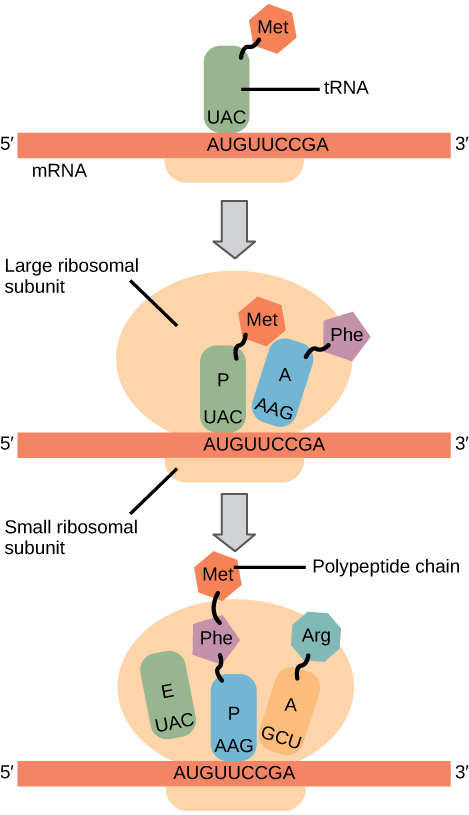
Many antibiotics inhibit bacterial protein synthesis. For example, tetracycline blocks the A site on the bacterial ribosome, and chloramphenicol blocks peptidyl transfer. What specific effect would you expect each of these antibiotics to have on protein synthesis?
Tetracycline would directly affect:
- tRNA binding to the ribosome
- ribosome assembly
- growth of the protein chain
Chloramphenicol would directly affect
- tRNA binding to the ribosome
- ribosome assembly
- growth of the protein chain
Termination of translation occurs when a nonsense codon (UAA, UAG, or UGA) is encountered. Upon aligning with the A site, these nonsense codons are recognized by release factors in prokaryotes and eukaryotes that instruct peptidyl transferase to add a water molecule to the carboxyl end of the P-site amino acid. This reaction forces the P-site amino acid to detach from its tRNA, and the newly made protein is released. The small and large ribosomal subunits dissociate from the mRNA and from each other; they are recruited almost immediately into another translation initiation complex. After many ribosomes have completed translation, the mRNA is degraded so the nucleotides can be reused in another transcription reaction.
Protein Folding, Modification, and Targeting
During and after translation, individual amino acids may be chemically modified, signal sequences may be appended, and the new protein “folds” into a distinct three-dimensional structure as a result of intramolecular interactions. A signal sequence is a short tail of amino acids that directs a protein to a specific cellular compartment. These sequences at the amino end or the carboxyl end of the protein can be thought of as the protein’s “train ticket” to its ultimate destination. Other cellular factors recognize each signal sequence and help transport the protein from the cytoplasm to its correct compartment. For instance, a specific sequence at the amino terminus will direct a protein to the mitochondria or chloroplasts (in plants). Once the protein reaches its cellular destination, the signal sequence is usually clipped off.
Many proteins fold spontaneously, but some proteins require helper molecules, called chaperones, to prevent them from aggregating during the complicated process of folding. Even if a protein is properly specified by its corresponding mRNA, it could take on a completely dysfunctional shape if abnormal temperature or pH conditions prevent it from folding correctly.
Summary
The players in translation include the mRNA template, ribosomes, tRNAs, and various enzymatic factors. The small ribosomal subunit forms on the mRNA template either at the Shine-Dalgarno sequence (prokaryotes) or the 5' cap (eukaryotes). Translation begins at the initiating AUG on the mRNA, specifying methionine. The formation of peptide bonds occurs between sequential amino acids specified by the mRNA template according to the genetic code. Charged tRNAs enter the ribosomal A site, and their amino acid bonds with the amino acid at the P site. The entire mRNA is translated in three-nucleotide “steps” of the ribosome. When a nonsense codon is encountered, a release factor binds and dissociates the components and frees the new protein. Folding of the protein occurs during and after translation.
Art Connections
Figure \(\PageIndex{2}\): Many antibiotics inhibit bacterial protein synthesis. For example, tetracycline blocks the A site on the bacterial ribosome, and chloramphenicol blocks peptidyl transfer. What specific effect would you expect each of these antibiotics to have on protein synthesis?
Tetracycline would directly affect:
- tRNA binding to the ribosome
- ribosome assembly
- growth of the protein chain
Chloramphenicol would directly affect
- tRNA binding to the ribosome
- ribosome assembly
- growth of the protein chain
- Answer
-
Tetracycline: a; Chloramphenicol: c.
Glossary
- aminoacyl tRNA synthetase
- enzyme that “charges” tRNA molecules by catalyzing a bond between the tRNA and a corresponding amino acid
- initiator tRNA
- in prokaryotes, called \(\text{tRNA}_\text{f}^\text{Met}\); in eukaryotes, called tRNAi; a tRNA that interacts with a start codon, binds directly to the ribosome P site, and links to a special methionine to begin a polypeptide chain
- Kozak’s rules
- determines the correct initiation AUG in a eukaryotic mRNA; the following consensus sequence must appear around the AUG: 5’-GCC(purine)CCAUGG-3’; the bolded bases are most important
- peptidyl transferase
- RNA-based enzyme that is integrated into the 50S ribosomal subunit and catalyzes the formation of peptide bonds
- polysome
- mRNA molecule simultaneously being translated by many ribosomes all going in the same direction
- Shine-Dalgarno sequence
- (AGGAGG); initiates prokaryotic translation by interacting with rRNA molecules comprising the 30S ribosome
- signal sequence
- short tail of amino acids that directs a protein to a specific cellular compartment
- start codon
- AUG (or rarely, GUG) on an mRNA from which translation begins; always specifies methionine